Renaissance Imaging Center Westlake Village
Saturday, December 21, 2019
14 Y?O with months history of knee buckling and swelling
Renaissance Imaging Center Westlake Village
Friday, December 20, 2019
35M runner with chronic hip pain MRI 2016 and Now
Thursday, December 19, 2019
Saturday, December 14, 2019
46F chronic ankle pain
![]() |
| What is your differential diagnosis? |
Pathology reference
Open access pediatric reference of MRI "flip-flop" effect in anorexia nervosa
Wednesday, December 11, 2019
31F professional dancer, collided with another dancer weeks ago. Persistent volar pain when making a fist.
![]() |
| Red arrow: retracted RCL tear. Yellow arrow: extensor mechanism injury |
![]() |
| Red arrow: retracted RCL tear |
![]() |
| You can see the thickened retracted torn RCL at the level of the SF MC head |
![]() |
| Bright signal localizes to oblique fiber tear of extensor mechanism. Orthopedist suspected volar plate injury; the volar plate is intact. |
Tuesday, December 10, 2019
![]() |
| Baseline available SIJ MRI 11 years ago at age 28 |
![]() |
| SIJ MRI 2014, only T1w and T2FSE non fat sat images done |
![]() |
| 1 year ago I reported chronic bilateral sacroilitis with questionable minimal synovial edema |
Monday, December 9, 2019
![]() |
| What would your DDx be? Is this neoplastic, infectious or inflammatory? What would you biopsy? |
![]() |
| Can leukemia / lymphoma infiltrate the tendon sheath like this? Would you consider sarcoid? Other? |
![]() |
| Femoral lymphadenopathy |
![]() |
| Subcutaneous, intermuscular and intramuscular edema |
![]() |
| Adenopathy with subcutaneous and intramuscular edema |
Friday, November 29, 2019
RE: doubled crossed patellar tendon
Hi,
Here is the summary of my inquiry about the crossed-doubled patellar tendon.
5x confident about prior tendon translocation surgery (Roux-Goldthwait procedure)
2x favouring an anatomical variant, citing the below reference from Loizides A et al., 2017
Coincidentally, today I have received new clinical information that my patient was treated surgically in the 90s for repeated patellar dislocation. Bingo, case solved!
In fact, I now have serious doubts about the existence of a doubled crossed patellar tendon anatomic variant and I would like to open for discussion based on the following:
While I think there is consensus about patellar tendon variants, like the one published here:
The uncrossed-doubled patellar tendon: A novel imaging finding
Syed Mohammed Ali Haydar, Jonathan Miles, Rikin Hargunani
Radiol Case Rep. 2019 Dec; 14(12): 1509–1512. Published online 2019 Oct 18. doi: 10.1016/j.radcr.2019.09.029
I do have serious concerns that the crossed-doubled patellar tendon, as it is performed by the Roux-Goldthwait procedure, actually exists as a variant because there is only 1 (one!) publication on this finding and it is referenced over and over again. The patient in this case report actually had 2x knee surgery for 'hemangioma', when the patient was 15 y/o! I find that suspicious and I have serious concerns about some missing information.
Alexander Loizides, Carmelo Messina, Bernhard Glodny, Leonhard Gruber, Erich Brenner, Hannes Gruber, Benjamin Henninger
Surg Radiol Anat. 2017; 39(1): 111–114. Published online 2016 Jun 15. doi: 10.1007/s00276-016-1706-x
So with all of that said, I am really wondering, if we in this wonderful community of MSK experts, could find a consensus if a crossed-doubled patellar tendon actually exists as a variant or not? I am not aware of this variant being described in any anatomical textbook and I have not seen any cases published or posted cases in children which I find very suspicious. Please correct me if I am wrong.
I am neither bored, nor looking for trouble but if we agree this specific anatomic variant does not exist, I question if this case report can or should stand any longer because it confuses people who search for this and always come up with the same paper!
BTW, this topic had been brought up previously on OCAD on Feb. 27th 2019 by Avneesh and others, also posting at least two other cases of doubled patellar tendon without known prior surgery. At least one case also had patellar malalignment! The colleagues are cc'd.
Modified Roux-Goldthwait Procedure for Management of Patellar Dislocation in Skeletally Immature Patients with Down Syndrome
https://www.ncbi.nlm.nih.gov/pmc/articles/PMC6394190/
Enjoy RSNA!
Björn
From: ocad-msk@googlegroups.com <ocad-msk@googlegroups.com> On Behalf Of Björn Jobke
Sent: 25 November 2019 15:52
To: OCAD <ocad-msk@googlegroups.com>
Subject: doubled crossed patellar tendon
---EXTERNAL EMAIL. Please exercise caution when opening attachments or clicking on links---
Hello OCAD,
My mystery case of the year.
52 y/o pat.
Relevant Clinical History: Recurrent patellar dislocation, since giving way whilst standing. Had injection for her pain few years back. Her pain has come back and bothering her too much. Diffuse tenderness present
No other history available.
No X-ray.
http://www.claripacs.com/a.php?a=125&mode=simple
What's going on? Variant or unknown prior surgical procedure?
Björn
--
Visit OCAD at xrayhead.com/ocad
---
You received this message because you are subscribed to the Google Groups "OCAD MSK" group.
To unsubscribe from this group and stop receiving emails from it, send an email to ocad-msk+unsubscribe@googlegroups.com.
To view this discussion on the web visit https://groups.google.com/d/msgid/ocad-msk/AM5P190MB0356C469C1B76409B6CD33A3A04A0%40AM5P190MB0356.EURP190.PROD.OUTLOOK.COM.
Wednesday, November 27, 2019
Happy Thanksgiving!
![]() |
| Here's my question, what is the Red Arrow pointing to? |
That's the terminal plantar fascia. In the forefoot, deep distal fibers insert onto the plantar plate, but superficial fibers insert onto the dermis. That's interesting, isn't it?
Tuesday, November 26, 2019
![]() |
| No history of trauma, acute onset of pain, reported as subhcondral fracture. |
![]() |
| Reported as normal |
![]() |
| Reported as normal |
![]() |
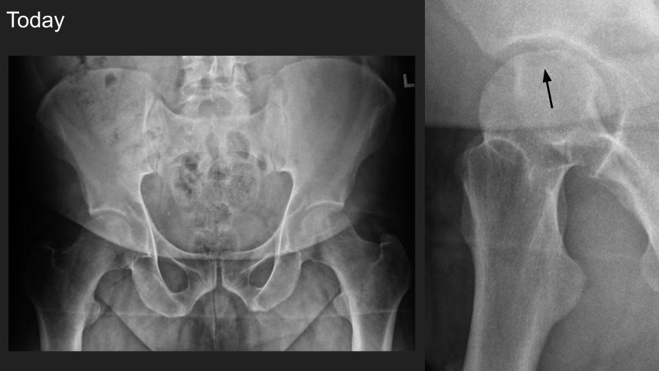
| There is new subtle smooth articular flattening visible in the Dunn view only |
Monday, November 25, 2019
Hypermobile discoid Lateral Meniscus in 9yo M with 2 mos posterior knee pain
![]() |
| There is posterocentral subluxation of the complete discoid LM with bright intrasubstance signal and expansion of the posterior horn region, but no visible surface defect. Wrisberg (which doesn't look thick) inserts onto the PH region. |
![]() |
| I cannot see popliteomeniscal fascicles, which I presume are developmentally absent. |
![]() |
OCAD LIVE 2020 Registration is Now Open
Join us for OCAD Live 2020! Saturday, March 28th 10 am - 5 pm UCLA - Santa Monica Orthopaedic Hospital Click HERE to register
-
A serious recreational runner with focal left shin pain and a lump after a long run is sent for ultrasound evaluation. Power doppler and gra...
-
F 62, left hip pain. Suspected impingement. There's a subchondral / subcortical bone lesion at the anterior aspect of the left femoral h...